DMA Measurements for Structure Determination of Anisotropic Fillers
Introduction
In polymer nanocomposites, inorganic particles are so finely distributed in the polymer matrix that at least in one dimension structures smaller than 100 nm can occur [1].
Well-known, classical nanofillers such as carbon black are isotropic. In contrast, sheet silicates such as montmorillonite are anisotropic and have a high length-to-thickness ratio (aspect ratio) of about 1:100. Besides the conventional composite materials, one distinguishes between two types of lamellar nanocomposites
In intercalated structures, the polymer chains alternate with the silicate layers in a fixed compositional ratio and have a well-defined number of polymer layers in the intralamellar space. In exfoliated nanocomposites, the number of polymer chains between the layers is almost continuously variable and the layers are largely delaminated.
The explanation of structure-property relationships based on the underlying morphology and filler structure is an important aspect in the development of nanocomposites.
Established techniques for structure determination include transmission electron microscopy (TEM) and small-angle X-ray scattering (SAXS). The spatial distribution of the exfoliated silicate layers can only be studied by TEM although SAXS can measure the separation of layers down to 1–4 nm. TEM is a rather complex method that allows qualitative information to be obtained about the internal structures and the spatial distribution of platelets. SAXS can only be used for intercalated structures, but not for exfoliated.
To investigate orientation effects of the local filer-filler-network, the materials used in this study were measured in different spatial directions by Dynamic Mechanical Analysis (DMA). Classical methods such as TEM and SAXS support the results of the DMA analysis. The main advantage of DMA analysis is the easy preparation and handling of samples.
Experimental Details
This present study used nanocomposites with hydrogenated nitrile butadiene rubber, HNBR, as the matrix rubber and modified montmorillonite (MMT) as the nanofiller. For the accurate elucidation of the structure and arrangement of the fillers and the effects on the mechanical properties, reference mixtures were prepared that contained only the matrix material, the filler and the crosslinking agent. The name of the sample is HNBR/OLS-5. It contains 5 phr organic layered silicate (OLS).
Measurements and Results
Investigation of the filler orientation
To detect possible orientation of the filler network or the silicate platelets, samples for measurement were taken from different spatial directions (see Figure 1).
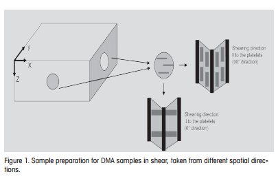
Due to the processing conditions (rolling in the x-direction), orientation of the exfoliated platelets is expected in the direction of flow.
Depending on the deformation direction, the storage moduli and tan d values from DMA investigations are different (see Figures 2 and 3). The modulus is higher parallel to the silicate layers (II) than at right angles (⊥). In comparison, the moduli of the unfilled references are lower and independent of the direction of deformation. The greater reinforcing effect in the II direction can be explained by assuming that the layers are arranged along the x-direction, which leads to an increase of the effective aspect ratio.
Comparable effects were found with polybutylene succinate (PBS). The marked increase of G′ (in comparison with other nanocomposites with the same clay concentration) can be explained by the formation of band-like structures [2]. This is probably the simplest model that can explain the properties of the material investigated in this study.
Morphology (TEM, SAXS)
To validate the DMA results, the filler dispersion was studied by transmission emission microscopy (TEM) using ultrafine sections. The TEM images of the HNBR/OLS-5 are displayed in Figures 4 and 5 and show that the platelets are exfoliated and oriented.
 |
DMA Measurements for Structure Determination of Anisotropic Fillers | Thermal Analysis Application No. UC 274 | Application published in METTLER TOLEDO Thermal Analysis UserCom 27